Monday, March 23, 2015

Spontaneous Retroperitoneal Hemorrhage

A middle aged male patient, was undergoing hemodialysis ( with anticoagulation), developed sudden drop of Hemoglobin 10 to 6g/dL.


Ultrasound scan showed right retroperitoneal hemorrhage. CT images showed a large right sided retroperitoneal hematoma, measuring ~10cm x12.5cm x 13cm.


Another smaller intramuscular hematoma was also seen along the right Iliacus muscle, reaching upto it's insertion.


Diabetic patients on hemodialysis are susceptible to develop spontaneous retroperitoneal hemorrhage. 'In patients receiving anticoagulant therapy in whom progressive anemia and unstable vital signs are present, spontaneous retroperitoneal bleeding should be considered as a possible cause.'[1].

'The presenting common symptoms and signs of retroperitoneal bleeding included sudden and progressive abdominal pain with blood pressure drop and subsequent development of an abdominal mass. These symptoms were associated with a falling hematocrit without any documented external blood loss and with suggestive X-ray changes, including absence of psoas shadow with soft tissue density.[2]

References :
[1] :  Spontaneous retroperitoneal bleeding: a case series : Hitoshi Yamamura. Sep 2014.
[2] : Ann Intern Med. 1977 Feb;86(2):189-92. Spontaneous retroperitoneal bleeding in patients on chronic hemodialysis.

Saturday, March 21, 2015

Orbital Blow-out Fractures

The CLASSIC BLOWOUT FRACTURE involves the floor of the orbit, usually sparing the orbital rim. Frequently, orbital tissues are trapped in the fracture site and ocular motility disturbances have been ascribed to entrapment of one or both inferior EOMs, but may be also due to orbital hematoma / inflammation.

The term orbital blow-out fracture describes the injury that results from a blow to the orbit by an object that is too large to enter the orbit (fist, baseball, etc.). The force of the blow is absorbed by the orbital rim and is transmitted to the thinner orbital floor, which shatters, usually in the middle third near the infraorbital canal. As the eye is pushed back into the conical orbital apex, it increases intraorbital pressure and this ‘‘blows out’’ the fractured floor into the maxillary sinus.

Pure blow-out Vs. Impure blow-out fractures
Usually the orbital rim is not fractured (pure blow-out fracture) and the globe remains undamaged. Less commonly the inferior orbital rim also is fractured; this is referred to as an impure blow-out fracture

Herniation of orbital fat, inferior rectus muscle, and inferior oblique muscle can occur with occasional muscle entrapment in the fracture line, resulting in diplopia on upward gaze.

Diplopia is the most frequent complaint in all patients with blow-out fractures and may occur solely because of periorbital edema and hemorrhage, which exert pressure on the globe. This type of diplopia resolves in several days, whereas entrapment diplopia remains.
Ref : Head and Neck Imaging, 4th Edition, Peter.N.Som.

Terms 'Blow-out / Blow-in' fractures can also be used in cases of Medial wall or rarely roof fractures also. Medial wall fractures are more likely to cause diplopia than a floor blow-out. Roof fractures might require an intracranial approach of treatment because of the breach of dura.


Sometimes the fractured floor, can sprang back into place, after the inferior rectus has herniated, trapping the muscle, this is called as a 'Trapdoor Fracture' (See the image below)


CT coronal section (soft tissue window) showing the entrapped inferior rectus, with the fractured floor back into anatomical position -- TRAPDOOR fracture.

Ref : RG 2006; 26:783-793 - 'Diagnosis of Midface fractures with CT'.

 
The extra-ocular muscle that herniates in the floor blow-out fracure is almost always the Inferior Rectus - Oblique. But below is a rare case, where the medial rectus has herniated through the floor defect into the maxillary sinus.

Click on the image to view in full-size


Friday, August 15, 2014

Active arterial extravasation in Blunt Abdominal Trauma


H/o Road Traffic Accident, Middle aged male, with maintained SpO2 and Blood Pressure.

The following plain CT sections show Hematoma involving right diaphragmatic crus with marked enlargement compared to the contralateral side. Hyperdensities s/o blood clots are noted within the hematoma. Mild amount of bilateral retroperitoneal hemorrhage was seen, with associated mild perinephric hemorrhage.


The following 3 images, shows the serial axial section during PLAIN / Non-contrast CT, Arterial Phase (25sec) and during Portal Venous Phase (60secs) respectively. Images demonstrate small area of contrast extravasation from the lateral margin of abdominal aorta, with minimal increase in the portal phase --> s/o an Active Arterial Hemorrhage.


These serial 5 images that follow, shows pooling of contrast in the 5 minute delayed images. Areas of contrast pooling are marked with large red arrows.


There was also a small anterior cortical laceration of right kidney.


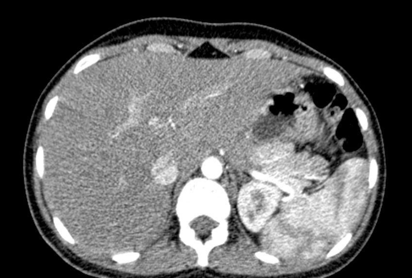
Pseudolesion : Papillary Process of Caudate Lobe


Here the papillary process is seen as an exophytic projection, seen extending into the perigastric region.
In the axial sections it appears as a separate 'lesion', in close relation to tail of pancreas.

In all imaging series, it is seen isoattenuating to the liver parenchyma.

CT for this young female patient was done for suspicion of appendicitis and renal calculi.

"The papillary process of the caudate lobe of the liver may appear separate from the liver and thus mimic lymph nodes or a pancreatic mass."  (Radiology. 1989 Dec;173(3):631-3. Papillary process of the caudate lobe of the liver: sonographic appearance. Donoso L1, Martínez-Noguera A, Zidan A, Lora F.)


LinkWithin

Related Posts Plugin for WordPress, Blogger...